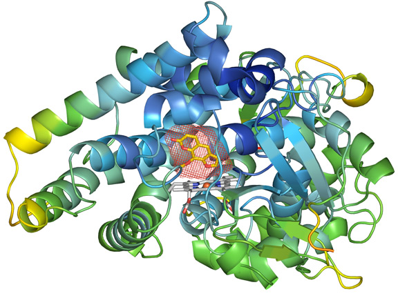
 The secondary structure representation of CYP2A6 with methoxsalen bound in the active site. The active site cavity (rendered as a thin red mesh) is just above the heme group, which is in the active site of the molecule. The inhibitory molecule, methoxsalen (yellow and red
stick figure), complements the size and shape of the cavity. Helices are represented as spirals, and beta-strands are represented as arrows. Oxygen atoms are colored red, nitrogen atoms blue and carbons are white for the heme group, and yellow for methoxsalen. Image by Jason Yano.
The secondary structure representation of CYP2A6 with methoxsalen bound in the active site. The active site cavity (rendered as a thin red mesh) is just above the heme group, which is in the active site of the molecule. The inhibitory molecule, methoxsalen (yellow and red
stick figure), complements the size and shape of the cavity. Helices are represented as spirals, and beta-strands are represented as arrows. Oxygen atoms are colored red, nitrogen atoms blue and carbons are white for the heme group, and yellow for methoxsalen. Image by Jason Yano.