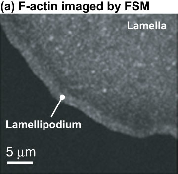
Fluorescent speckle microscopy, as shown in this image, is based on a method developed by Associate Professor Clare Waterman-Storer and her former boss E.D. Salmon. This recent technique helps to tease apart the mechanics of cell movement by tracing how the actin cytoskeleton undergoes continuous flow from the leading edge toward the cell body. Image by Aaron Ponti.


The actin network that propels the cell forward is actually composed of two distinct, overlapping networks of actin polymers assembling at the leading edge of a cell. This image reveals these two distinct populations. Fast, short-lived speckles (shown in red) cluster in the lamellipodium, whereas slow, long-living speckles (shown in black) populate the lamella, but expand all the way to the leading edge, revealing a spatial overlap of two distinct actin networks. Image by Aaron Ponti.