| |
Vol. 4 Issue 22 / July 19, 2004
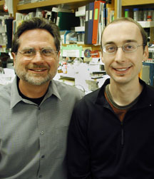
Structure Shows How One Human Protein Reduces the Potency of ChemotherapyA team of researchers led by scientists at The Skaggs Institute for Chemical Biology at The Scripps Research Institute have solved the structure of a human protein called AGT that is known to interfere with the action of certain chemotherapy drugs. AGT repairs damaged DNA inside human cells. Cancer cells can use it to repair DNA that has been damaged in the course of chemotherapy—thus rendering the chemotherapy ineffective. The structure of AGT, which is being reported in an upcoming issue of the journal Nature Structural & Molecular Biology, was solved in the laboratory of Scripps Research Professor John Tainer by Research Associate Douglas Daniels. The fine details of this x-ray structure may help scientists find ways to fight certain cancers, such as brain tumors, which express high levels of AGT making them resistant to chemotherapy. "This structure provides a solid foundation for the development of better drugs and resistant proteins [intended to improve cancer therapies]," says Tainer. Cancer, Chemotherapy, and ResistanceCancer is one of the greatest public health scourges in the United States today. According to the U.S. Centers for Disease Control and Prevention, it is the second leading cause of death. In 2001, the most recent year for which such statistics are available, 553,768 people died of some form of cancer. For years, one of the best available ways to treat cancer has been through the administration of chemotherapy drugs, such as "alkylating" agents, which inhibit the growth of cancer cells by damaging their DNA. DNA, the molecule that stores our genetic information, is composed of two intertwining helical strands that bind to each other through base pairing. Each strand is composed of four types of DNA bases (A, G, C, and T) attached to one another through a sugar-phosphate backbone. The other strand in the pair has a complementary sequence of bases. Alkylating agents are chemicals that transfer one of a number of different carbon-chain "alkyl" groups onto the bases of DNA, which change the structure of those bases and often disrupt the transcription or replication of that DNA. This can retard a cell's growth and lead to its death—the desirable outcome for cancer cells. The damage these agents cause may not be confined to cancer cells, but the toxicity to the cancer cells is usually greater than the toxicity to the rest of the body. However, the human repair protein O6-alkylguanine-DNA alkyltransferase (AGT), which is one of the body's natural defenses against DNA damage, can inadvertently protect cancer cells from these agents. AGT is remarkably efficient and repairs DNA extremely rapidly, says Anthony Pegg of the Pennsylvania State University College of Medicine, who has been studying AGT protein for several years and is one of the authors on the study. However, says Daniels, this protection can cut both ways. "Tumor cell lines hijack [AGT's] function and overexpress the protein to escape therapies," he says. Cells in some tissues, such as the brain, naturally have high levels of AGT. This enables cancers that arise in the brain to resist chemotherapy. Cancer cells in other parts of the body can also use AGT to escape chemotherapy by "upregulating" or expressing excess amounts of AGT. When these cancers do this, they are rendered resistant to alkylating agents. Wanting to understand how AGT repair works on the molecular level, Daniels and Tainer teamed up with Pegg a few years ago to solve the structure of AGT. Twist and GrabNow Daniels and Tainer have solved the structure of AGT bound to DNA. The structure gives a view of how the protein binds to damaged DNA, says Pegg, and this information is useful for designing inhibitors to interfere with AGT's actions. In fact, Pegg has already made such inhibitors of AGT, and some of these are currently being tested in clinical trials as an adjuvant for chemotherapy. The idea is that these inhibitors could be administered at the same time as chemotherapy to make chemotherapy more effective. "Having the structure solved allows us to make [inhibitors] that are [even] more selective," Pegg says. The structure also provides a way of designing proteins to improve cancer treatments, says Tainer. The molecular details of the structure show how AGT could be modified to resist selective inhibitors. This could be used to protect critical tissues from the chemotherapy even when administered at high doses. For example, a cancer patient taking chemotherapy and AGT inhibitors could at the same time receive an infusion of modified bone marrow cells that would have a form of AGT protein able to resist the inhibitors, which could protect the patient from some of chemotherapy's more toxic side effects. Daniels and Tainer had already solved the free-form structure of AGT (not bound to DNA), and when they compared this to the DNA-bound form, they found that the structures were identical. The fact that AGT's structure does not change after binding the DNA suggests a simple one-step pathway whereby AGT repairs damaged DNA. The model that Daniels, Tainer, Pegg, and their colleagues are suggesting is one of twist and grab. AGT recognizes a piece of DNA that is damaged and binds to that DNA. Then it twists out the phosphate backbone of the DNA, exposing the damaged base, repairs the base by grabbing the offending alkyl group and transferring it onto itself, and releases the now repaired section of DNA. In doing so, the protein is used up. DNA commonly twists in such a way that there is a major groove (like the part of the staircase with the steps) and a minor groove (like the part with the rail). One of the details that has emerged from this study is that AGT recognizes the minor groove of DNA. Most proteins that bind to DNA bind in the major groove. Interestingly, major groove binders have the same structural motif as AGT, which binds in the minor groove. In fact, says Tainer, "One third of all DNA-binding proteins use this same motif to bind DNA in the major groove." The article, "DNA binding and nucleotide flipping by the human DNA repair protein AGT" by Douglas S. Daniels, Tammy T. Woo, Kieu X. Luu, David M. Noll, Neil D. Clarke, Anthony E. Pegg, and John A. Tainer is an advance online publication of the journal Nature Structural & Molecular Biology and may be accessed at: http://dx.doi.org/10.1038/nsmb791. The article will appear in print in the August 2004 issue of the same journal. This work was funded by the National Institutes of Health, the National Cancer Institute, the Patterson Trust, the Alexander and Margaret Stewart Trust and The Skaggs Institute for Research.
Send comments to: jasonb@scripps.edu
|
|
|