| |
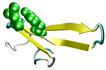
Folding, Misfolding, and Lessons from a WW DomainProtein misfolding can be such a difficult and abstract subject that it becomes easy to lose sight of the simple principles that scientists would like to understand regarding how and why proteins misfold. One reason can be illustrated by the discovery a doctor made almost 100 years ago. When Dr. Alois Alzheimer examined a post-mortem patient who died with an unusual mental illness in 1906, he found clumps of "amyloid" protein plaques in her brain. These plaques are still a clear sign of the disease that bears his name—a disease now believed to inflict some 4 million Americans. Many diseases are caused by the formation of protein plaques inside the body. Amyloid-forming diseases like Alzheimer's and Parkinson's are well known, but others include a collection of over 80 rare amyloid diseases caused by the misfolding of the protein transthyretin (TTR), which the liver secretes into the bloodstream to carry thyroid hormone and vitamin A. In diseases like familial amyloid polyneuropathy (FAP), hundreds of these proteins misfold into structures leading to microscopic fibril plaques, which deposit in internal organs and interfere with normal function, sometimes lethally. Another reason for scientists' interest in protein misfolding can be illustrated by a problem recently facing a graduate student. The student was toiling away at a routine laboratory procedure of expressing and purifying a small soluble protein in bacteria, but every time the student induced the expression of the protein, the protein molecules would clump together on the nanoscale, form insoluble "inclusion bodies" on the microscale, and crash out of solution like a fried egg at the bottom of the test tube on the macroscale. In fact, understanding protein misfolding would have many applications in basic biochemistry, where it could be used to prevent such aggregation and advance basic laboratory methods. As such, says Jeffery W. Kelly, who is the Lita Annenberg Hazen Professor of Chemistry at The Scripps Research Institute (TSRI) and vice president of academic affairs at TSRI, understanding how and why proteins misfold is a high priority for many scientists. And, he adds, "In order to completely understand misfolding, we have to understand how proper protein folding takes place." A Model For Beta Sheet FormationNow two studies in an upcoming issue of Proceedings of the National Academy of Sciences (PNAS) provide insight into the folding process of what are known as beta sheet structures. This is a common fold or "motif" wherein fully extended peptide strands hydrogen bond to each other to form a sheet—the same way that plastic teeth in a flat comb line up. In one article, Kelly, his colleagues at The Skaggs Institute for Chemical Biology at TSRI, and a group at the University of Illinois examined the kinetics of beta sheet formation using a protein fragment called the formin-binding protein (FBP) WW domain. The "WW" refers to the fact that there are two conserved tryptophan residues in the hundreds of sequences comprising this domain family (tryptophan is abbreviated "W"). This FBP WW domain is a small protein fragment of a few dozen residues that folds rapidly into its structure in tens of microseconds. The structure is very basic—its chain folds to form three strands of a beta sheet connected by two loops. Significantly, this structure is incredibly tolerant to mutagenesis—two sequence-dissimilar WW domains from divergent species will nevertheless fold into the same three-dimensional structure. This tolerance is important to be able to mutate residues of this protein and gauge which ones may be particularly important for the folding of these WW domains. And that's exactly what Kelly and his colleagues did. They made changes to the protein, including mutating certain residues, cutting off the end of the FBP WW domains, and observing protein folding under temperature variations. They observed this by monitoring a change in fluorescence as the proteins adopts or changes its structure using a laser heating and rapid fluorescence measurement technique employed by the University of Illinois group. What the researchers found was that temperature, mutation, and truncation changes all have the ability to alter the kinetics of the protein folding—changing the pathway by which these proteins fold, not their final structure. The folding kinetics or the way that FBP and other WW domain proteins fold can generally be divided into two separate classes—the two-state folders and the three-state folders. Two-state folders form the characteristic beta sheet in a single transition. That is, they go from one (unfolded) state to a second (folded) state. Three-state folders, on the other hand, go through an intermediate state, which means that they have two transitions along their folding pathways (unfolded to intermediate and intermediate to folded). Kelly and his colleagues found that when the FBP WW domains folded with three-state kinetics, the formation of a particular loop between two individual beta strands is the rate-limiting step for the folding of the beta sheet, meaning that these connections form more slowly. They also found that it is possible to "tune" the way the proteins folded by changing the sequence or the temperature of the protein. Specifically, Kelly and his colleagues were able to switch a slow folding, two-transition WW domain into a more rapid, single transition WW domain. Kelly explains that higher temperatures, the removal of the C-terminus of the protein, or cetain internal mutations destabilize the intermediate state, so the proteins fold in a single transition. A Model of the FoldingA second PNAS paper, also scheduled for publication soon, by TSRI graduate student John Karanicolas and Charles L. Brooks III, professor of molecular biology at TSRI, predict this behavior with a new model. Karanicolas and Brooks model the folding kinetics of two different WW domains—which both fold into the classic WW beta sheet, but via the two different mechanisms. Significantly, Karanicolas and Brooks found through their simulations that the origin of the three-state folding is a particular set of residues that form mismatched contacts with each other in the intermediate state. These contacts occur during the formation of a particular loop that must form before the beta strands can line up to form a beta sheet. This process has the propensity to form a misaligned loop in which the incorrect neighbors line up, which is sort of like mismatching the buttons on your shirt. This mismatched intermediate, Karanicolas and Brooks found, is what drives the rate-limiting step. They observed this misaligned loop and inferred that one particular residue that is responsible for fixing the alignment is removed, the slow phase gets slower. Karanicolas and Brooks shared this result with Kelly, and Kelly and his colleagues made mutations to the proteins, tested their folding, and verified the prediction experimentally. What emerges from both of these studies is a complicated description best explained by a simple analogy: the folding of the WW domain is something like buttoning up your shirt in the morning. If you misalign the buttons, it takes time to unbutton the shirt and button it up again. In other words, it's faster to button your shirt right the first time than to button it incorrectly and have to fix it, and it's possible to "tune" the process by adjusting surrounding conditions, like the light in the room. Turn up the dimmer, and you can see the shirt better. The better you see, the less likely you are to make a mistake buttoning your shirt, and the faster you will be able to do it. "This [research] was an extraordinarily good set of interactions," says Brooks. "Jeff [Kelly] and his colleagues were really listening to our theoretical input, and we were getting all this wonderful feedback that helped us understand our models better and led, ultimately, to the correct picture of the folding for this protein." To read the article "Tuning the free energy landscape of a WW domain by temperature, mutation, and truncation" by Houbi Nguyen, Marcus Jager, Alessandro Moretto, Martin Gruebele, and Jeffery W. Kelly, please see: http://www.pnas.org/cgi/doi/10.1073/pnas.0538054100. To read the article "The structural basis for biphasic kinetics in the folding of the formin-binding protein WW domain: Lessons for protein design?" by John Karanicolas and Charles L. Brooks III, please see: http://www.pnas.org/cgi/doi/10.1073/pnas.0731771100.
|
| |