| |
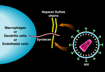
Attachment Receptors and Hot Spots for HIV InfectionIn the world of human immunodeficiency virus (HIV) research, much of the focus for the last couple of decades has been on the host receptors that are necessary for the entry of HIV into the cell—the CD4 receptor and its co-receptor CCR5, for instance, which HIV uses to enter helper T cells. So much attention has been given to these CD4 receptors, in fact, that they have become almost synonymous with HIV itself. Loss of CD4 receptors has long been a defining diagnostic of AIDS, and the level of CD4 cells is a marker of disease progression. However, this is only the start of the story. There are also "attachment" receptors, which have been shown in recent years to enhance the entry of HIV into cells. "Now more than ever, there is a growing body of knowledge that suggests attachment receptors can have a profound impact on HIV pathogenesis," says Assistant Professor Philippe Gallay, who is a member of the Department of Immunology at The Scripps Research Institute (TSRI). In his laboratory, Gallay looks at the attachment of the virus to cells and looks toward using those host proteins as a guide for drug design. Hooked on SugarsThe particular class of attachment receptor that interests Gallay consists of long, extended proteins on the surface of human cells that are decorated with a kind of sugar known as heparin sulfate chains. Heparin sulfate chains are attached to "glyco" proteins on the surface of cells like macrophages, which use them for a number of biological reasons, like binding to cytokines and growth factors in the bloodstream. Gallay and his colleagues showed a few years ago that these heparin sulfate chains are also important players in the pathology of HIV because cells that are decorated with these heparin sulfate chains are like glue for the virus. Heparin sulfate chains have affinity for the viral coat protein GP120 on HIV, and HIV seems to use them to gain entry into cells like macrophages, one of the virus's main target cells. "When you remove these sugars, the virus cannot infect macrophages," says Gallay, who demonstrated this a few years ago in a study with Research Associate Andrew C. S. Saphire. But the sugars are only part of the story. Recently, Gallay and members of his laboratory published a paper describing the primary importance of a human protein called syndecan, which contains certain "motifs" of amino acids that the body's heparin sulfate chains attach to, which in turn interact with HIV's viral coat protein GP120. The group that published this finding included Michael D. Bobardt, Saphire, Hsiu-Cheng Hung, and Xiaocong Yu at TSRI and their colleagues Bernadette Van der Schueren, Zhe Zhang, and Guido David of the Center for Human Genetics at the University of Leuven and Flanders Interuniversity Institute in Belgium. HIV CoOpts the Machinery of the CellSyndecans are actually a family of four different highly conserved transmembrane proteins that sit on the outside of cells. While no syndecan structures have been solved yet, their extracellular domains are known to extend from the cell surface, and at their terminus, they usually have motifs to which heparin sulfate chains are covalently attached. Syndecans are the connection between the cells and the extracellular matrix, the molecular scaffold the body uses to build collections of cells into tissues. The interaction of syndecans with extracellular matrix components induces signals inside cells that are related to adhesion and migration. Cells that are adhering to the extracellular matrix express high levels of syndecans, while T cells and other mobile cells that do not adhere to the extracellular matrix do not express syndecans. "They [Syndecans] probably have several biological roles," says Gallay, "such as presenting cytokines and growth factors to receptors." Significantly, this machinery that binds to cytokines and growth factors in the bloodstream is what HIV has hijacked for its own purposes. It tricks the syndecans into capturing virions rather than growth factors. "We found that syndecans can capture a lot of virus via their long extended chains," says Gallay. He had previously shown that syndecans are expressed on the surface of macrophages and that, by removing the syndecans, they were able protect the macrophages from infection. He also showed that monocytes, the precursor cells from which macrophages are derived, do not express syndecans and cannot be infected with HIV, whereas macrophages do express syndecans and can be infected. So syndecans, because of the affinity of their heparin sulfate chains for GP120, can be used by HIV to gain entry into cells. But there is more. In his recent study, published in the January 2003 issue of the journal Immunity, Gallay and his colleagues showed that syndecans could act as in trans receptors, meaning that cells decorated with syndecans not only capture HIV, they help it to infect other cells that do not express the syndecans. In other words, the presence of syndecans on one cell can help HIV enter another cell.
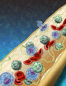
The capture of virus by syndecans and their role as in trans receptors has had a profound impact on how Gallay and others think about HIV pathogenesis. In his most recent study, Gallay and his colleagues found that various subtypes of HIV, including HIV-1 and HIV-2, and even related lentiviruses, like simian immunodeficiency virus (SIV) use syndecans as in trans receptors for entry. "Syndecans not only capture HIV—they protect it as well," says Gallay. Normally, free virus loses its ability to infect cells relatively quickly in the bloodstream as it is exposed to proteases, chemicals, and components of the immune system. But while free virions lose their activity after a single day, those bound to the long chains of the syndecans on the outside of cells are protected from the proteases and other blood components, and remain infectious for up to a week. During this week, syndecans can present the virions to passing CD4+ T cells, potentially infecting them. Moreover, Gallay and his colleagues showed that the syndecans not only present the virions, they actually enhance their infectivity because they concentrate the virus into certain anatomical hot spots. This is especially significant because syndecans are broadly expressed by the body's cells in the inside surface of blood vessels and other tissue similarly lined with endothelial cells. Endothelial cells are one of the major cell types of the body, accounting for about one percent of the total cells in the body—approximately 600 square meters of surface area. "We found that all of the endothelium is rich in these syndecans," says Gallay. Significantly, endothelial cells that line the adenoids, tubing that connects the lymph nodes to the blood stream, are literally covered with syndecans—meaning that they may also be covered in virus. This is very important, says Gallay, because as T cells go in and out of the lymph nodes, they may be picking up the virus. Now he is attempting to generate with phage display antibodies that target the part of the HIV GP120 coat protein that interacts with the heparin sulfate chains. This will not only help him identify the particular domains of GP120 to which the heparin sulfate chains bind, but the antibodies that might interrupt the interaction of syndecans with HIV, preventing attachment of HIV to these cells. "The final goal is to prevent the virus from being protected by the endothelial wall and see if the virus will be rapidly degraded," says Gallay. Significantly, such antibodies might also be relevant as a starting point for prophylactic protection against HIV. Gallay notes that syndecans are particularly richly expressed on genital epithelial cells, where they can rapidly accumulate the virus—something that may be relevant for the initial transmission of the virus into the bloodstream. "I think the best place [to attempt to] block transmission of the virus is in this initial stage," says Gallay. "We hope to find drugs or antibodies that disrupt transmission into the blood stream."
|
| |