The Ins and Outs of Endocytosis
By Jason Socrates Bardi
The easiest way to describe endocytosis is to think not of cells but
of sports arenas—crowded with star players, role players, benchwarmers,
waterboys, coaches, referees, and spectators, and with lots of ticket
holders wanting to come in.
Endocytosis is like an extremely efficient V.I.P. entrance to the arena:
an usher gathers a group of important people at one of the gates. Then
the gate is pulled inward, enveloping the people and becoming an elevator
car that whisks them away to their skybox. The elevator car then returns
the ushers back to the stadium where they can gather more ticket holders.
To speak less metaphorically, receptor-mediated endocytosis is an essential
cellular process whereby important hormones, proteins, nutrients, and
other macromolecular "cargo" needed by a cell are collected and transported
across plasma membranes—those lipid bilayers that define the outer
edges of eukaryotic cells.
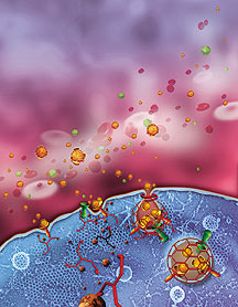
Receptors (ushers) gather proteins and other nutrients (spectators)
and pinch them off in a membrane packet surrounded by a protein cage.
Scientists refer to these packets as "coated vesicles," and the machinery
that forms them and regulates their formation is complex, involving numerous
structural proteins and accessory factors. A cell biologist's dream.
"We're interested in all aspects of how that machinery functions," says
Sandra Schmid, chair of the Department of Cell Biology.
Coated Vesicles
Endocytosis is essential for development, when it plays a key role in
carting cascades of molecules that establish the gradients necessary for
stem and other precursor cells to develop into specialized cells and tissues.
While cells with mutations that render them unable to undergo endocytosis
can survive in culture, the same mutations are always lethal to organisms.
Endocytosis also plays a larger role in biology, providing cells in
a mature organism with a way to take in essential molecules from the bloodstream.
Insulin and cholesterol, for instance, are transported into cells through
receptor-mediated endocytosis.
Endocytosis is also important for medical reasons because toxins and
viruses co-opt the machinery to gain entry into cells. In contrast, the
process may provide a vehicle for transporting beneficial drugs into cells.
The Receptors and the Cages
The receptors that mediate endocytosis are large proteins that span
the membranes of cells. The outside, "extracellular" portion has binding
sites that allow it to catch molecules of interest outside the cell. The
inside, "intracellular" portion carries an address label or "sorting signal"
that is its ticket into the endocytic vesicle. These two regions of the
receptor are connected through a "transmembrane" portion so
that binding of cargo to the outside can be sensed to activated sorting
signals on the inside.
"Cargo molecules and their receptors aren't just passengers. They can
also be active drivers in this process controlling the type of vehicle
they take, the speed at which it travels and the final destination," says
Schmid.
The receptors also take cues from the cell. To maximize the efficiency
of receptor-mediated endocytosis, the cell instructs the receptors to
gather and concentrate in a certain region of the membrane so that they
can, in turn, gather and concentrate the molecules of interest in that
small patch of membrane outside the cell. And while the receptors are
gathering the cargo on the outside of the cell, other molecules on the
inside of the cell are busy making a "vesicle" container to transport
it in.
Vesicles are actually just patches of membrane where receptors are and
where the cargo molecules are being gathered. That patch of membrane becomes
involuted, bulging inward to form a pit that is surrounded on the inside
of the cell by a lattice-like coat of protein known as clathrin. (Endocytosis
is also sometimes referred to as clathrin-mediated endocytosis in recognition
of this protein's essential role.)
The clathrin surrounds the involuting patch of membrane, which then
pinches off to form the tiny vesicles. One way to envision the process
is to imagine yourself in the fruit and vegetable aisle of the grocery
store. Receptor-mediated endocytosis is like putting your hand inside
a plastic bag, grabbing a bunch of green beans, and then turning the bag
inside out around them.
Although clathrin is the primary scaffold of the protein cage, clathrin
does not go it alone. Other proteins are also involved in the formation
of the coated vesicle. The assembly of the clathrin coat is controlled
by other regulatory elements of the cell—such as the regulatory protein
dynamin.
"Dynamin is central to the process of clathrin-mediated endocytosis,"
says Schmid, and she points to fruit fly (Drosophila) mutants discovered
three decades ago as proof. These particular mutants have expressed a
wounded dynamin protein that is active at low temperatures but inactive
at high temperatures. The flies are fine at low temperature, but at high
temperature the mutation is lethal and causes cells to lose their ability
to carry out endocytosis. As a result of this loss of function, the flies
never fully develop.
The mutation, as it turns out, is in the dynamin gene—the same
gene that Schmid's laboratory first identified over a decade ago and that
she has been studying ever since.
Conan the Dynamin
Dynamin is responsible for finally pinching off the "neck" of the budding
vesicle, which releases it into the interior of the cell. Dynamin's control
of this essential final step led Schmid to recently refer to the enzyme
as a master regulator of the late stages of vesicle formation.
Interestingly, dynamin is actually several enzymes in one. At one end,
its amino terminus, dynamin has a GTPase domain—a portion that when
folded correctly can "hydrolyze" or clip off a phosphate group from a
GTP molecule.
Dynamin also carries some of its own activating proteins. Normally,
GTPase enzymes require cofactor proteins (called "GAP" for GTPase-activating
protein) to be active. Dynamin carries its own GAP.
As far as GTPases go, dynamin is something of a standout. Small GTPases
average around 20,000 Daltons, and large ones are something like 40,000
Daltons. Dynamin is about 100,000 Daltons—a protein chain of over
800 amino acids.
"It's the Arnold Schwarzenegger of GTPases," says Schmid.
In 1995, Schmid found that dynamin self-assembles at the neck of budding
vesicles, which led her to propose the first model for how dynamin works.
According to this model, the dynamin self-assembles at the neck of the
forming vesicle and pinches it off, freeing the vesicle to traffic through
the cell. In referring to this mechanism, she likened the action of dynamin
to the assassin's murder weapon the garrote—it tightens around the
neck, and pop.
In subsequent years, Schmid's laboratory went on to probe this mechanism
of action in greater detail, concentrating on how the dynamin self-assembles
and how it tightens around a budding vesicle. The whole time she was looking
for evidence to support this model.
"In fact," she says, "We found evidence that the model was wrong."
Dynamin, Schmid now believes, is a much more sophisticated molecule.
"Dynamin is not just the brawn," Schmid says. "It's part of the brains."
She thinks that it is integrating the process of endocytosis with other
events in the cell. It may be rearranging the actin cytoskeleton of the
cell, monitoring what is entering cell-wide, and inducing a stress response.
A Powerful Assay
The biochemical assay that the Schmid laboratory developed involves
purifying plasma membranes, stripping them of all their materials, and
reconstituting the machinery to generate coated vesicles and recreate
each of the steps leading to endocytosis in the test tube. This assay
has allowed her to study and understand the action of dynamin and to identify
the other cellular machinery that carries out vesicle formation.
"It is different than assays we have used in the past, which use a single
receptor and a single ligand," says Schmid. "Now we can look at any receptor
we want."
One of the things these studies have revealed is that the regulation
of receptor-mediated endocytosis is a highly sophisticated interaction
between the cargo molecules, the receptors, dynamin and other enzymes,
and the clathrin coat.
In addition to having binding sites outside the cell that recognize
and collect the cargo molecules, receptors have binding sites on their
portions inside the cell that are used for binding as well—to the
clathrin molecules that form the cage around the budding vesicle.
However, clathrin does not recognize the receptors directly. The cell
employs "adaptor" proteins that recognize the sorting motifs on the receptors
and then "adapt" the cargo molecules to the coat. Like the familiar three-pronged
to two-pronged converters people use to plug their toaster ovens into
old outlets, adaptor molecules fit the receptors to the clathrin scaffold
during vesicle formation.
Moreover, the adaptors exert control over endocytosis because they trigger
assembly of the clathrin coat, and they group the receptors together,
thus concentrating the cargo. And to make the situation even more elaborate,
the adaptors are, in turn, controlled by other parts of the cellular machinery.
About a year ago, the Schmid laboratory discovered a new "kinase" enzyme
that is involved in the regulation of cargo selection by controlling the
adaptors. The kinase that Schmid found attaches a phosphate group to adaptor
molecules and thereby regulates cargo selection by altering the adaptor.
The adaptor molecule is called AP-2, and Schmid's new kinase binds to
it and attaches a phosphate group to the portion of AP-2 that is responsible
for recognizing the receptor molecule. Schmid found that when the kinase
attaches the phosphate to the adaptor molecule the affinity for the cargo
molecules increases 25-fold.
"That's a huge difference," she says. "Kinases usually have a 2- to
3-fold effect."
Into the Cell
However, when Schmid and her colleagues went beyond the biochemical
assay and designed a series of mutant cells that would allow them to see
what happens when they tinker with the kinase, they were surprised.
"We learned things we never could have anticipated from what we had
done in the test tube," she says.
What they anticipated was that by overexpressing the kinase, they would
be overphosphorylating the AP-2 adaptor molecules. They reasoned that
the overphosphorylated adaptors would be randomly distributed and unable
to cluster.
Then, since adaptor clustering leads to clathrin clustering, Schmid
and her laboratory thought that overexpressing the kinase would shut down
vesicle formation and endocytosis.
However it did not.
In fact, to their surprise, they found that the clathrin distribution
was not affected by essentially knocking out the function of AP-2. Other
adaptors, they concluded may be working independently of AP-2 to accomplish
the same goal.
"AP-2 may be just another cargo-specific adaptor," she concludes.
In general, she adds, receptor-mediated endocytosis seems to be more
sophisticatedly regulated than was ever previously thought.
"In retrospect this makes sense," says Schmid. "Cells communicate between
themselves and with their environment through the plasma membrane. Endocytosis
plays a critical role in regulating this communication."
Go back to News & Views Index
|