Nucleotide-Dependent Motor Movement
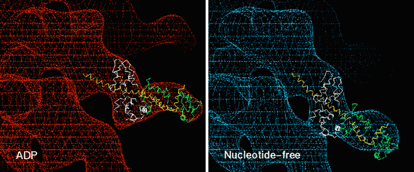
Figure 3. 3D maps (wire frame) of smooth muscle myosin II attached to F-actin in the presence of Mg-ADP (Left) and with no nucleotide (rigor) (Right). No change in orientation of the motor domain is seen when ADP is released, but the light chain domain swings ~23°. Docking the chicken skeletal x-ray structure of the light chain domain into the EM maps suggests that this domain swings as a rigid body on ADP release (see
Whittaker et al. 1995).
Return to the Actomyosin Cryo-Electron Microscopy
contents © 2000
awl